Knee Conditions

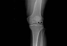
Knee Fracture
A fracture is a condition in which there is a break in the continuity of the bone. In younger individuals, these fractures are caused from high energy injuries, as from a motor vehicle accident. In older people, the most common cause is weak and fragile bone
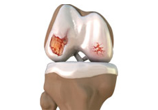
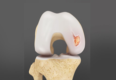
Knee Osteoarthritis
Osteoarthritis, also called degenerative joint disease, is the most common form of arthritis. It occurs most often in older people. This disease affects the tissue covering the ends of bones in a joint (cartilage). In a person with osteoarthritis, the cartilage becomes damaged and worn out causing pain, swelling, stiffness and restricted movement in the affected joint.
Ligament Injuries
- Anterior Cruciate Ligament (ACL)
- Medial Collateral Ligament (MCL)
- Posterior Cruciate Ligament (PCL)
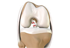
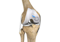
ACL Tears
An ACL injury is a sports-related injury that occurs when the knee is forcefully twisted or hyperextended. An ACL tear usually occurs with an abrupt directional change with the foot fixed on the ground or when the deceleration force crosses the knee. Changing direction rapidly, stopping suddenly, slowing down while running, landing from a jump incorrectly, and direct contact or collision, such as a football tackle can also cause injury to the ACL.
ACL Sprain
The anterior cruciate ligament, or ACL, is one of the major ligaments of the knee that is in the middle of the knee and runs from the femur (thighbone) to the tibia (shinbone). It prevents the tibia from sliding out in front of the femur. Together with posterior cruciate ligament (PCL) it provides rotational stability to the knee.

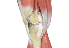
MCL Tears
Medial collateral ligament (MCL) injury can result in a stretch, partial tear, or complete tear of the ligament. Injuries to the MCL commonly occur because of pressure or stress on the outside part of the knee. Anterior cruciate ligament (ACL) may be torn along with an MCL injury.
MCL Sprain
The medial collateral ligament (MCL), a band of tissue present on the inside of your knee joint, connects your thighbone and shinbone (bone of your lower leg). The MCL maintains the integrity of the knee joint and prevents it from bending inward.
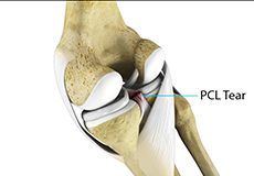
PCL Injuries
PCL injuries are very rare and are more difficult to detect than other knee ligament injuries. Cartilage injuries, bone bruises, and ligament injuries often occur in combination with PCL injuries. Injuries to the PCL can be graded as I, II or III depending on the severity of injury. In grade I, the ligament is mildly damaged and slightly stretched, but the knee joint is stable.
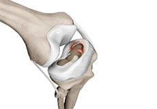
Meniscal Injuries
Meniscal tears often occur during sports. These tears are usually caused by twisting motion or over flexing of the knee joint. Athletes who play sports such as football, tennis and basketball are at a higher risk of developing meniscal tears. They often occur along with injuries to the anterior cruciate ligament, a ligament that crosses from the femur (thighbone) to the tibia (shinbone).
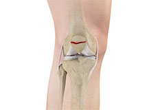
Meniscal Tears
Meniscus tear is the commonest knee injury in athletes, especially those involved in contact sports. A sudden bend or twist in your knee causes the meniscus to tear. This is a traumatic meniscus tear. Elderly people are more prone to degenerative meniscal tears as the cartilage wears out and weakens with age.
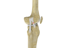
Patellar Fracture
The kneecap or patellar is the largest sesamoid bone in the body and one of the components of the knee joint, present at the front of the knee. The undersurface of the kneecap and the lower end of the femur are coated with articular cartilage, which helps in smooth movement of the knee joint. The kneecap protects the knee and provides attachment to various muscle groups of the thigh and leg.
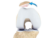
Patellar Dislocation
Dislocation of the patellar occurs when the patellar moves out of the patellofemoral groove, (called as trochlea) onto a bony head of the femur. If the kneecap partially comes out of the groove, it is called as subluxation and if the kneecap completely comes out, it is called as dislocation (luxation).
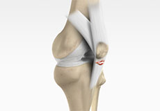
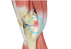
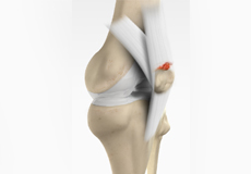
Patellar Tendon Rupture
Patellar tendon rupture is the rupture of the tendon that connects the patellar (kneecap) to the top portion of the tibia (shinbone). The patellar tendon works together with the quadriceps muscle and the quadriceps tendon to allow your knee to straighten out.
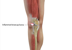
Lateral Patellar Compression Syndrome
Lateral patellar compression syndrome refers to pain under and around your kneecap. It is a common complaint among runners, jumpers, and other athletes such as skiers, cyclists and soccer players.
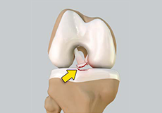
Tibial Eminence Fractures
The tibia or shin bone is a major bone of the leg which connects the knee to the ankle. A fracture or break in the upper part of the tibia is known as a proximal tibial fracture and commonly occurs just below the knee joint.
Osgood Schlatter Disease
Osgood-Schlatter disease refers to a condition of an overuse injury that occurs in the knee region of growing children and adolescents. This is caused by inflammation of the tendon located below the kneecap (Patellar tendon). Children and adolescents who participate in sports such as soccer, gymnastics, basketball and distance running are at higher risk of this disease.
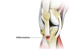
Jumper’s Knee
Jumper’s knee, also known as “Patellar tendinitis" is an inflammation of the patellar tendon that connects your kneecap (Patellar) to your shinbone. This tendon helps in extension of the lower leg.
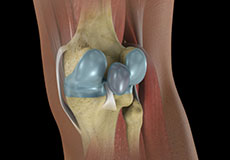
Baker’s Cyst
The knee consists of a fluid called synovial fluid, which reduces friction between the bones of the knee joint while you move your leg. Sometimes this fluid is produced in excess, resulting in its accumulation in the back of your knee. A Baker’s cyst or popliteal cyst is a fluid-filled swelling that develops into a lump behind the knee.
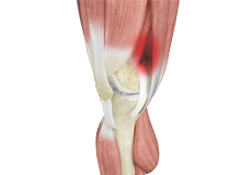
Iliotibial Band Syndrome
Iliotibial band syndrome is an overuse injury resulting from the inflammation of the iliotibial band. Iliotibial band is a tough group of fibres that begins at the iliac crest of the hip and runs along the outside of the thigh, to get attached to the outer side of the shinbone just below the knee joint. Its function is to coordinate with the thigh muscles and provide stability to the knee joint.
Osteochondritis Dissecans
Osteochondritis dissecans is a joint condition in which a piece of cartilage, along with a thin layer of the bone separates from the end of the bone because of inadequate blood supply. The separated fragments are sometimes called “joint mice”. These fragments may be localized, or may detach and fall into the joint space causing pain and joint instability.
Articular Cartilage Defects
Articular cartilage does not have a direct blood supply to it, so has less capacity to repair itself. Once the cartilage is torn it will not heal easily and can lead to degeneration of the articular surface, leading to development of osteoarthritis.
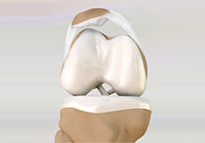
Quadriceps Tendon Rupture
Surgery is performed on an outpatient basis cannot be repaired arthroscopically since the tendon is outside the joint. The goal of the surgery is to re-attach the torn tendon to kneecap and to restore the normal function of the knee. Sutures are placed in the torn tendon which is then passed through the holes drilled in the kneecap.
Osteonecrosis of the Knee
Osteonecrosis is a condition in which death of a section of bone occurs because of lack of blood supply to it. It is one of the most common causes of knee pain in older women. Women over 60 years of age are commonly affected, three times more often than men.
Knee Angular Deformities
Angular deformities of the knee are common during childhood and usually are variations in the normal growth pattern. Angular deformity of the knee is a part of normal growth and development during early childhood. Physiologic angular deformities vary with age as: